ULTRASOUND
ULTRASOUND
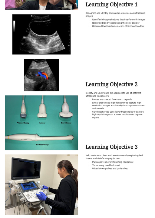
During the ultrasound portion of the program, students gained a foundational understanding of how to recognize and interpret anatomical structures on ultrasound images. They observed scans of the lower abdomen, identifying key organs such as the liver and bladder, and learned how to spot common imaging challenges—like ribcage shadows that can interfere with clarity. Using color Doppler, students also observed blood flow within vessels, gaining insight into how ultrasound is used to evaluate circulation and internal function in real time.

STUDENT WEBSITE
Students were introduced to different types of ultrasound transducers and their specific clinical uses. They learned that probes are made from quartz crystals and use varying frequencies depending on the exam. For example, the linear probe produces high-resolution images at shallow depths, making it ideal for viewing muscles and blood vessels, while the curvilinear probe offers deeper imaging for organs but at a lower resolution. In addition to image interpretation and equipment knowledge, students also practiced maintaining a clean and safe work environment by replacing bed sheets, disinfecting probes and beds, and following proper hygiene protocols—reinforcing the importance of infection control and professionalism in clinical settings.