
MAGNETIC
RESONANCE IMAGING
ABOUT THE EXPERIENCE
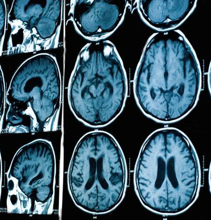
During the MRI portion of the program, students built a strong understanding of human anatomy by learning to read and interpret MRI images. They identified key structures such as the upper and lower abdominal organs and the cranial cavity, and observed how these areas appear when IV contrast (dye) is used to enhance visibility. This hands-on learning experience helped students see how MRI is used to support accurate diagnosis and treatment planning. They also gained insight into how different tissues and organs present on scans, building both technical knowledge and confidence in identifying anatomical features.
.png)
A major focus of this module was understanding MRI equipment and safety. Students learned about the different types of MRI coils—such as the breast, spine, and head coils—and how each one is designed for specific types of imaging. Most importantly, they were taught that the MRI magnet is ALWAYS ON, a critical safety concept in any MRI environment. Through observation and instruction, students learned how to read safety signs, follow proper room protocols, and ensure that non-magnetic items like jewelry are removed before entering MRI zones. This emphasis on safety gave students a real-world perspective on the importance of attention to detail, preparation, and professionalism in a clinical setting.
STUDENT WEBSITE
